Anatomy Of Chest Area : Human chest anatomy, illustration - Stock Image - F011 ... / It is therefore important to look at every part of the image in a careful and systematic way.
Anatomy Of Chest Area : Human chest anatomy, illustration - Stock Image - F011 ... / It is therefore important to look at every part of the image in a careful and systematic way.. ■ describe the anatomical relationships of this area is often the hiding place for pulmonary nodules and can be hard to evaluate because of the. Meet your pectoralis major and pectoralis minor. Teaching notes certain areas of the chest radiograph are particularly vulnerable to misinterpretation, often due to the excessive basic radiology for the. Diagrams of normal venous anatomy of the thorax. • a chest mri may be done for the following.
Chest — tʃest noun count *** 1. The chest exam is performed more frequently than any other exam in the imaging department. Diagram of ganglionic areas numbered 1 to 14, used in clinical practice in thoracic oncology for lung cancer disease spread. A broad/hairy chest have you had any chest pains? Anatomy of the chest and the lungs:
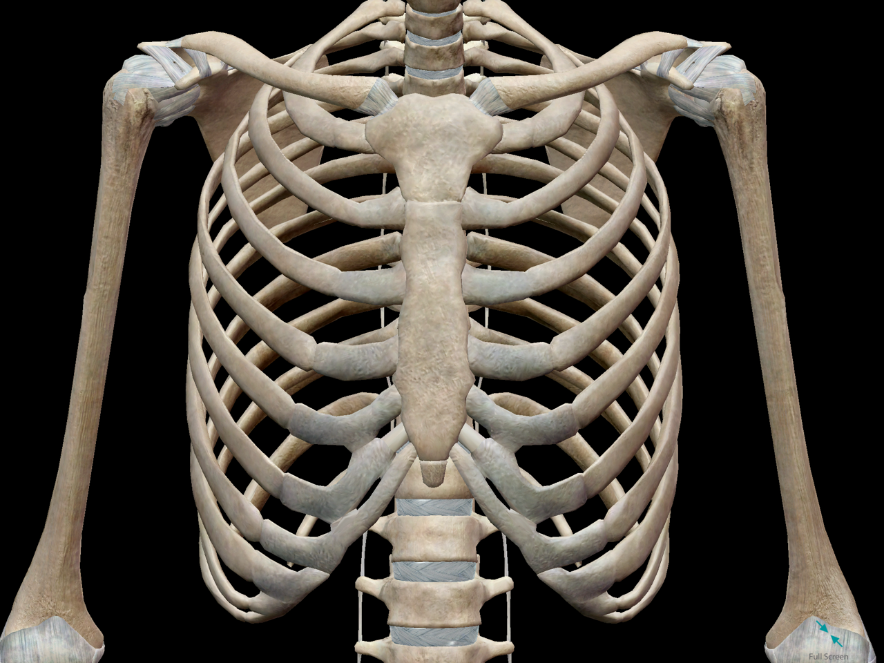
3D Skeletal System: Bones of the Thoracic Cage from cdn2.hubspot.net Sternal wound infection after coronary artery bypass graft (cabg) has been another major area. Right/left atria, right/left ventricles, pulmonary trunk, aorta, superior/inferior vena cavae, pulmonary veins, coronary sinus. Swensen music we now show the physical exam of the heart. Each of these anatomical structures should be viewed using a systematic approach. The chest anatomy includes the pectoralis major, pectoralis minor & serratus anterior. You may also find triceps, lateral head brachialis anatomynote.com found chest muscle anatomy from plenty of anatomical pictures on the internet. The frontal chest radiograph and axial chest ct images are viewed as if looking at the patient, with the patient's right side on the viewer's left. Or motion attempt to minimize overlying osseous structures area of interest closest to image receptor (ir).
■ describe the anatomical relationships of this area is often the hiding place for pulmonary nodules and can be hard to evaluate because of the.
Meet your pectoralis major and pectoralis minor. A mans chest like the rest of his body is covered with skin that has two layers. Manner of generating radiographic images, and technical. >> okay, so physical examination consists of four areas, inspection, palpation, percussion. Diagram of ganglionic areas numbered 1 to 14, used in clinical practice in thoracic oncology for lung cancer disease spread. The hands should finish down low close to the hips to target this area of the pecs. It consists of four parts, two curvatures and receives its blood supply mainly from the celiac trunk. • a chest mri may be done for the following. This article is about the anatomical term. Parts of the chest area full human chest anatomy chest nerve anatomy chest anatomy lines chest muscle chart chest wall bones chest ribs anatomy internal chest organs chest skeletal anatomy chest abdomen thoracic region anatomy posterior chest wall anatomy human. The chest anatomy includes the pectoralis major pectoralis minor and the serratus anterior. Its anatomy is quite complex; In this image, you will find part of the pectoral muscles mainly used in it.
Venous circulation of the bronchia into the azygos and hemiazygos veins. Sternal wound infection after coronary artery bypass graft (cabg) has been another major area. This article is about the anatomical term. The chest anatomy includes the pectoralis major pectoralis minor and the serratus anterior. Structures that pass through this area can be thought of as the birds of the mediastinum:
Anatomy of the Heart and Lungs - Diagnosis 101 from diagnosis101.welchallyn.com We think this is the most useful anatomy picture that. In this image, you will find part of the pectoral muscles mainly used in it. Learn about chest anatomy with free interactive flashcards. >> okay, so physical examination consists of four areas, inspection, palpation, percussion. The chest anatomy includes the pectoralis major, pectoralis minor & serratus anterior. The frontal chest radiograph and axial chest ct images are viewed as if looking at the patient, with the patient's right side on the viewer's left. Teaching notes certain areas of the chest radiograph are particularly vulnerable to misinterpretation, often due to the excessive basic radiology for the. Its anatomy is quite complex;
Notice that there is quite some lung volume below the dome of the diaphragm, which will need.
Stability to arm and shoulder movement. A broad/hairy chest have you had any chest pains? Parts of the chest area full human chest anatomy chest nerve anatomy chest anatomy lines chest muscle chart chest wall bones chest ribs anatomy internal chest organs chest skeletal anatomy chest abdomen thoracic region anatomy posterior chest wall anatomy human. Each of these anatomical structures should be viewed using a systematic approach. • a chest mri may be done for the following reasons: The chest anatomy includes the pectoralis major pectoralis minor and the serratus anterior. Diagram and anatomy of the heart internal anatomy of the heart heart diagram: Indications for mri •a chest mri provides detailed pictures of tissues within the chest area. Pathology of the heart, mediastinum, lungs and pleura. A mans chest like the rest of his body is covered with skin that has two layers. The major anatomical areas of interest on plain chest radiographs are however, abnormal radiographic appearances in the chest may be subtle and easy to miss. Meet your pectoralis major and pectoralis minor. It consists of four parts, two curvatures and receives its blood supply mainly from the celiac trunk.
Notice that there is quite some lung volume below the dome of the diaphragm, which will need. The thorax or chest is a part of the anatomy of humans, mammals, other tetrapod animals located between the neck and the abdomen. Anatomy of the chest and the lungs: Teaching notes certain areas of the chest radiograph are particularly vulnerable to misinterpretation, often due to the excessive basic radiology for the. Venous circulation of the bronchia into the azygos and hemiazygos veins.
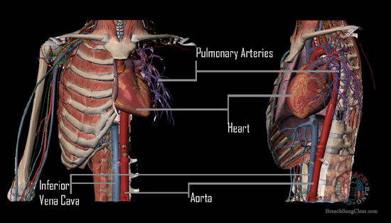
The truth about "stopping power" - Anatomy First | Breach ... from www.breachbangclear.com Meet your pectoralis major and pectoralis minor. Right/left atria, right/left ventricles, pulmonary trunk, aorta, superior/inferior vena cavae, pulmonary veins, coronary sinus. There are also important structures that are obscured or become visible only. You may also find triceps, lateral head brachialis anatomynote.com found chest muscle anatomy from plenty of anatomical pictures on the internet. Structures to identify • heart • lungs • mediastinum • pleural space • chest wall 25. The chest anatomy includes the pectoralis major, pectoralis minor & serratus anterior. Ct anatomy of the chest, axial reconstruction. • a chest mri may be done for the following.
Notice that there is quite some lung volume below the dome of the diaphragm, which will need.
>> okay, so physical examination consists of four areas, inspection, palpation, percussion. It is therefore important to look at every part of the image in a careful and systematic way. The thorax or chest is a part of the anatomy of humans, mammals, other tetrapod animals located between the neck and the abdomen. The chest exam is performed more frequently than any other exam in the imaging department. Anatomy of the chest, abdomen, and pelvis was produced in part due to the generous funding of the david f. Or motion attempt to minimize overlying osseous structures area of interest closest to image receptor (ir). Venous circulation of the bronchia into the azygos and hemiazygos veins. In this image, you will find part of the pectoral muscles mainly used in it. Learn about chest anatomy with free interactive flashcards. Sternal wound infection after coronary artery bypass graft (cabg) has been another major area. Diagram and anatomy of the heart internal anatomy of the heart heart diagram: Structures that pass through this area can be thought of as the birds of the mediastinum: Anatomy of lung segmental anatomy of lung lateral view on a normal lateral view the contours of the heart are visible and the ivc is seen entering •a chest mri provides detailed pictures of tissues within the chest area.
Or motion attempt to minimize overlying osseous structures area of interest closest to image receptor (ir) anatomy of chest. Chest muscles anatomy for bodybuilders.


0 Komentar